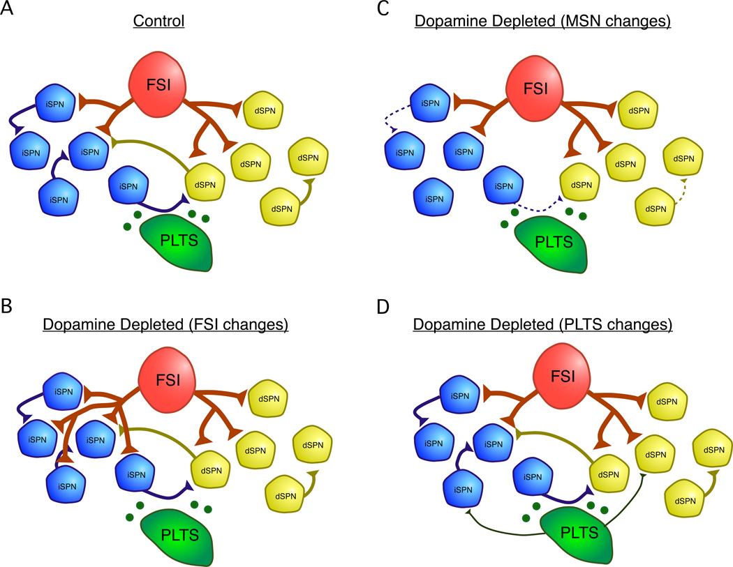
Figure 2.
Summary of changes in GABAergic microcircuits following dopamine depletion. A. Under normal conditions, SPNs laterally inhibit each other and inhibition is observed both across and within dSPN and iSPN subtypes 28. FSIs inhibit both dSPNs and iSPNs, but preferentially target dSPNs 8. PLTS interneurons release neuropeptides such as NPY and SOM and the neuromodulator NO which can modulate SPN activity 14. Under control conditions, inhibitory synapses from PLTS interneurons onto SPNs are hard to detect 8. The following changes have been observed to GABAergic microcircuits following pharmacological dopamine depletion in mice: B. Sprouting of FSI axons and the formation of new axons specifically onto iSPNs. This causes an inversion of the normal pathway-selectivity of FSIs such that after dopamine depletion, FSIs are more likely to target iSPNs than dSPNs 25. C. Reduction in connectivity and unitary strength of lateral inhibition between SPNs. Connections between dSPNs were sparse under control conditions and were no longer detected in dopamine depleted mice 28. D. An increase in the strength or connectivity of inhibitory inputs from PLTS interneurons onto SPNs may occur. This finding is based on increases in the frequency of large amplitude inhibitory postsynaptic currents (IPSCs) observed in SPNs after dopamine depletion 59, presumably arising from spontaneously active PLTS interneurons in the slice.

