
Fig. 9.
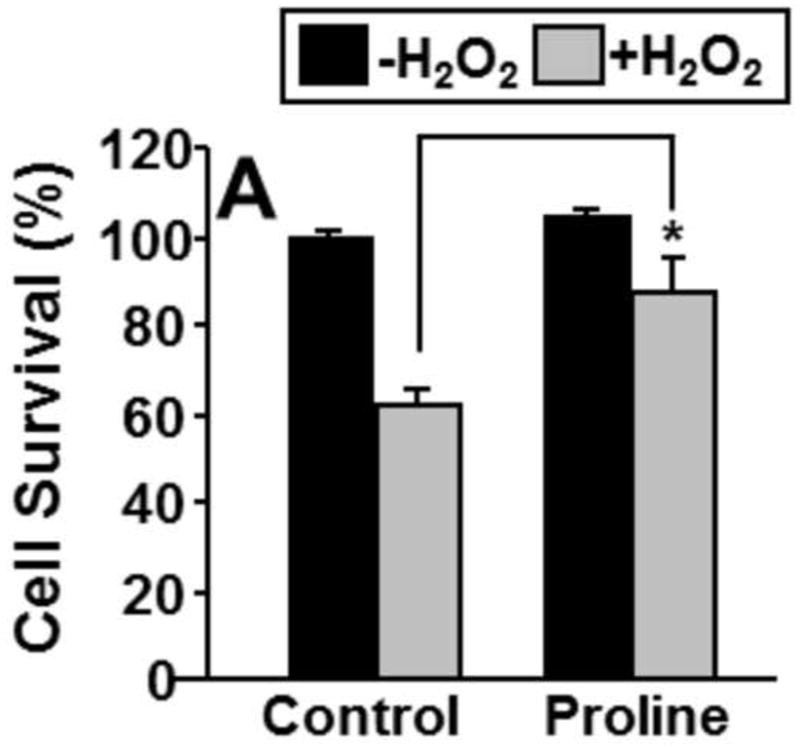
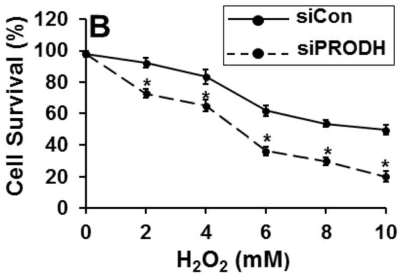
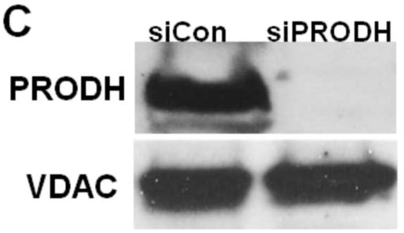
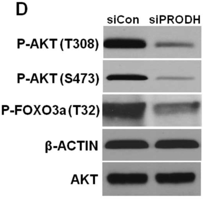

Knockdown of PRODH and Akt signaling pathway in prostate cancer cells. (A) RWPE-1 cells were treated with and without proline (5 mM) for 12 h and then incubated with 0.5 mM H2O2 for 3 h in serum free medium. Percent cell survival was estimated using the MTS cell viability assay. (B) Survival rates of PC3 cells transfected (48 h) with control siRNA (siCon) and PRODH siRNA (siPRODH) and incubated with different concentrations of H2O2 (0–10 mM) for 3 h. (C) Western blot analysis of PRODH in PC3 cells transfected with siCon (50 nM) and siPRODH (50 nM) for 48 h. VDAC is shown as a control. (D) Western blot analysis of P-Akt (T308 and S473), P-FoxO3a (T32), and β-actin in PC3 cells transfected with siCon and siPRODH (48 h). (E) Quantification of P-Akt-T308 and P-Akt-S473 relative to total AKT (P-Akt/Akt) and P- FoxO3a-T32 relative to β-actin. Each value represents mean ± SD of separate experiments (n = 4) (* P < 0.05).