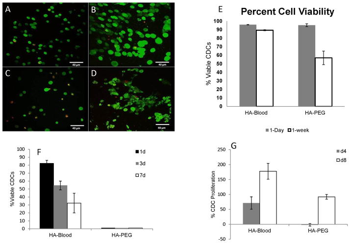
Figure 2. HA-blood hydrogels promote cell spreading and proliferation.
Representative 2-photon microscopy images illustrating live (green) and dead CDCs (red) on day 1 in HA-PEG (A) and HA-blood (human, lysed) hydrogels (B) and day 7 in HA-PEG (C) and HA-blood (D) hydrogels cultured in CEM
E. Bar graphs summarize CDC survival at 1d and 1wk in HA-blood and HA-PEG hydrogels cultured in CEM
F. HA-blood hydrogels, but not HA-PEG hydrogels permit CDC survival when cultured in Tyrode solution (containing glucose and electrolytes, but no serum)
G. Picogreen assay revealed CDC proliferation on d4 and d8 in HA-blood hydrogels, but only on d8 in HA-PEG hydrogels

