Figure 6.

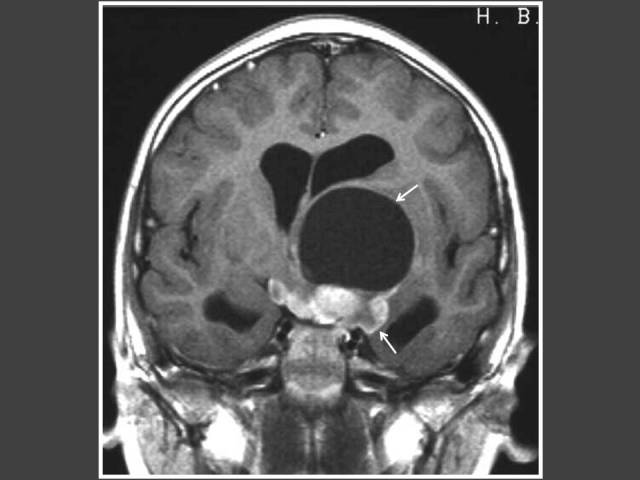
Four year-old boy who presented with impaired vision and acne. T1WI post-gadolinium sagittal MRI of the brain shows heterogenously enhancing suprasellar tumour (arrows) with hydrocephalus. Histology revealed pilomyxoid astrocytoma.

Four year-old boy who presented with impaired vision and acne. T1WI post-gadolinium sagittal MRI of the brain shows heterogenously enhancing suprasellar tumour (arrows) with hydrocephalus. Histology revealed pilomyxoid astrocytoma.