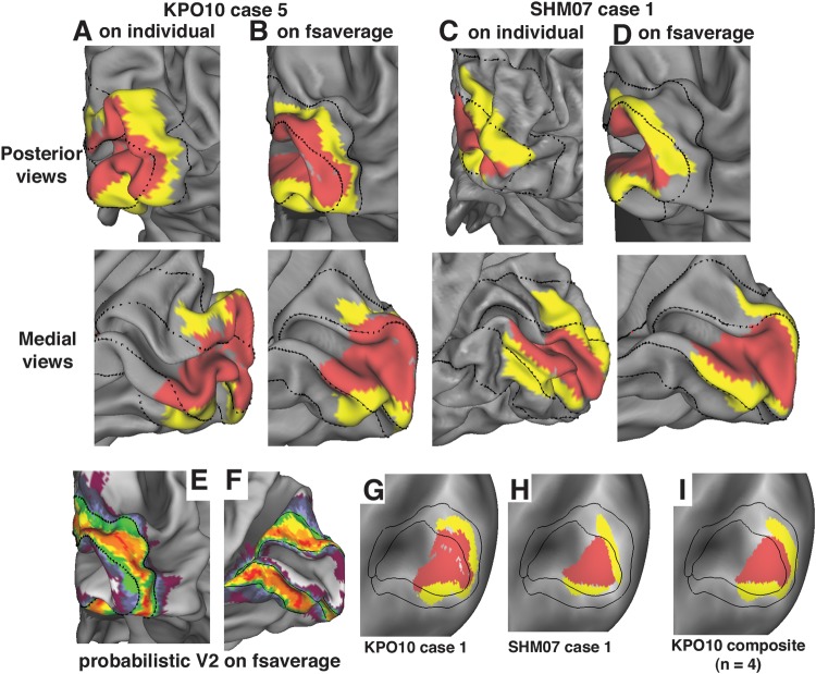
Figure 10.
Retinotopic areas V1 and V2 relative to probabilistic areas 17 and 18. (A) Map of retinotopic V1 and V2 from Case 1 of KHP10 (Kolster et al. 2010), displayed on the posterior (top) and medial (bottom) views of the right hemisphere midthickness surface, with architectonic area 17 and 18 boundary contours overlaid. (B) The same KHP10 case 1 retinotopic areas displayed on the fsaverage midthickness surface. Areal boundaries are in similar locations relative to the occipital pole and other geographic landmarks in the vicinity. (C,D) Retinotopically defined area V1 and V2 boundaries from case 1 of SHM07 (Swisher et al. 2007) displayed on the individual midthickness surface (C) and on the fsaverage midthickness surface (D). Areal boundaries are close to the occipital pole on both surfaces. (E,F). Probabilistic map of architectonic area 18 from Fischl et al. (2008) on posterior (E) and medial (F) views of the fsaverage midthickness surface. (G–I) Maps of retinotopic V1 and V2 of KHP10 case 1 (G), SHM07 case 1 (H), and KHP10 composite map (I). The eccentricity range spanned in these studies (7.75° for KHP10; 6–7.5° for SHM07) should have covered a little less than half of V1, based on previous studies in macaques (Van Essen et al. 1984) and humans (Hinds et al. 2009).

