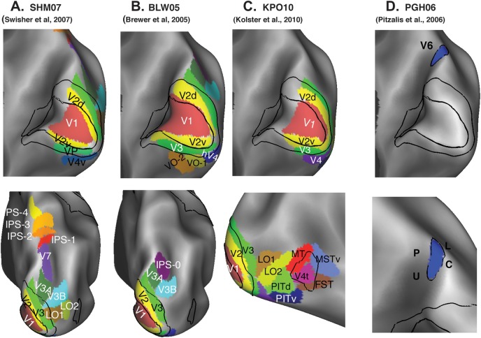
Figure 12.
(A) Retinotopic maps in human extrastriate cortex. A composite of the left and right hemisphere maps for one subject shown in medial (top) and dorsal-posterior views. (B) BLW05 (Brewer et al. 2005) retinotopic areas shown in the same views, the surfaces were smoother than a typical human anatomical surface. To improve registration to the PALS-B12 atlas, additional landmarks were added that were readily discernible in the individual and atlas surfaces. (C) KPO10 areas (Kolster et al. 2010). (D) Area V6 from PGH06 (Pitzalis et al. 2006).

