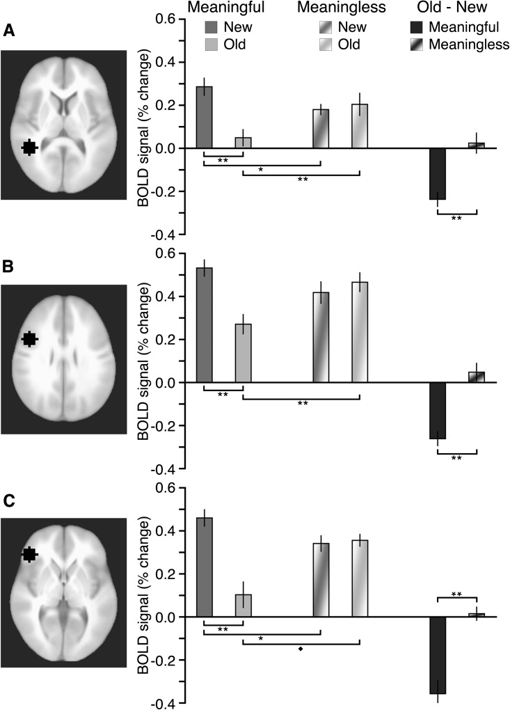
Figure 3.
Activity in conceptual brain regions in response to squiggles. Each of the 3 ROIs is shown at its middle axial slice, including: (A) left inferior temporal cortex, (B) left dorsal inferior frontal cortex, and (C) left ventral inferior frontal cortex. Estimated activity is shown for each ROI for meaningful and meaningless old and new squiggles, as well as for the old–new activity difference for meaningful and meaningless squiggles. Error bars indicate SE. All significant pairwise differences are highlighted. **P < 0.01. *P < 0.05. ♦P = 0.07.

