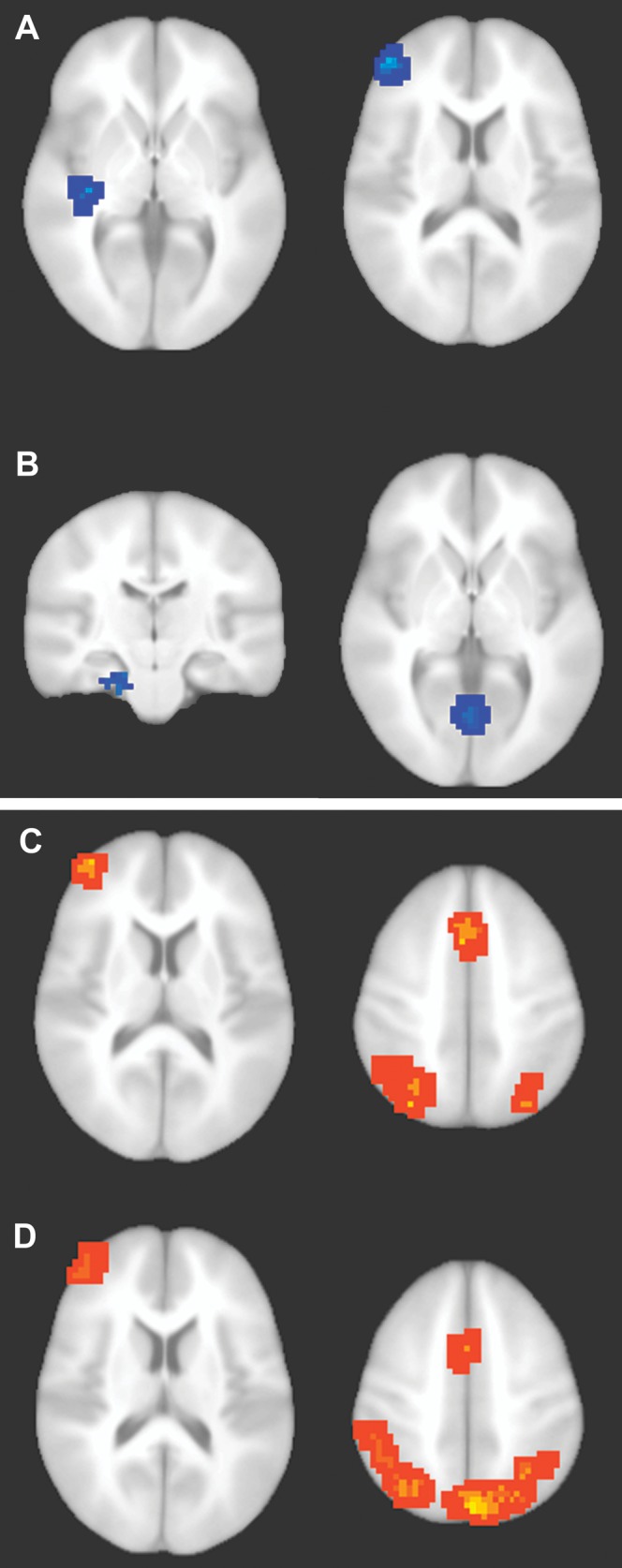
Figure 4.

Neural correlates of conceptual priming and recognition. (A–B) Regions associated with conceptual priming were identified by the double subtraction [meaningful (old − new) − (meaningless (old − new)]. Blue coloration indicates significantly greater activity reductions for meaningful squiggles than for meaningless squiggles. (A) Left inferior temporal cortex (left side) and left inferior frontal cortex (right side). (B) Left anterior entorhinal/perirhinal cortex (left side) and bilateral anterior lingual gyrus (right side). Conceptual priming clusters are described in Table 1. (C–D) Regions associated with recognition for meaningful and meaningless squiggles. (C) Hot coloration indicates regions showing a linear trend of significantly more activity across the remember, know, guess, and new response types for old meaningful squiggles (remember > know > guess > new). Findings were typical of prior studies of recognition memory using words and nameable pictures (e.g., Fig. 3 of Donaldson et al. 2001). (D) Regions showing the same linear trend for meaningless squiggles. Recognition clusters are described in Table 3.
