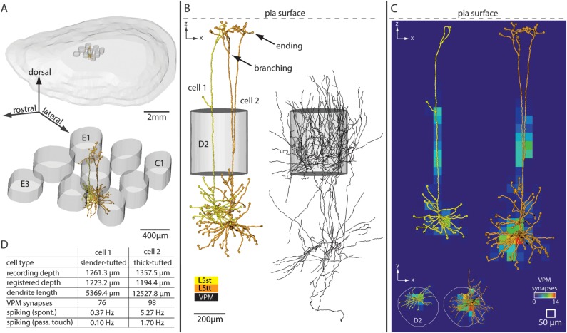
Figure 1.
Three-dimensional reconstruction and registration of in vivo-labeled dendrite and axon morphologies in a rat barrel column. (A) Upper panel: 3D view of pia surface and 9 L4 barrels. Lower panel: Blow up of the 9 barrels. The central barrel contains registered 3D soma–dendrite reconstructions. (B) Semicoronal view of registered neurons. Two excitatory neurons of different cell types are located at the same cortical depth (left) and are innervated by thalamocortical axons from VPM (right). (C) Structural overlap between registered dendrite and VPM axon morphologies allows determining 3D subcellular innervation of thalamocortical synapses. (D) This method of reconstruction and registration allows determining the cell type, anatomical parameters, synaptic innervation, and spiking activity in vivo (spontaneous and evoked by passive whisker touch) for individual neurons. This is illustrated for one example of 2 L5 pyramidal neurons shown in panels A–C.

