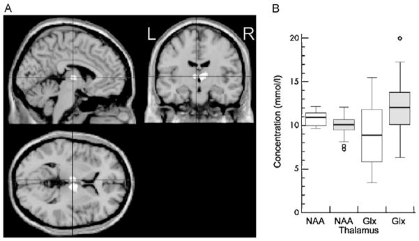
Fig. 5.
Decreased N-acetyl aspartate (NAA) and increased glutamate and glutamine (Glx) and reduced gray matter fraction in the thalamus of idiopathic generalized epilepsy (IGE) patients when compared with controls. (A) VBM showing decreased thalamus gray matter fraction in patients compared to controls. The white areas in the center of each image indicate z scores of ≥4. (B) Absolute concentrations of thalamic NAA and Glx in IGE (shaded) and controls (blank). The boxes indicate inner quartiles, the vertical lines the outer quartiles. Outliers are shown as circles.
This figure is reproduced from Helms et al. [44] with permission.

