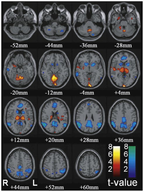
Fig. 6.
Increases in thalamus and decreases cortical regions are the most prominent changes with conventional HRF modeling in CAE patients. Functional data are superimposed on the MNI brain template “colin27” (sing subj T1 in SPM2) displaced in radiological right–left convention. In total, 54 seizures in nine patients were analyzed using GLM with canonical HRF in SPM2. fMRI increases were seen in bilateral thalamus, occipital cortex, and to a lesser extent the midline cerebellum, anterior and lateral temporal lobes, insula, and adjacent to the lateral ventricles. fMRI decreases were seen in the bilateral parietal, medial parietal, and cingulate cortex and basal ganglia.
This figure is reproduced from Bai et al. [5] with permission.

