Figure 3.

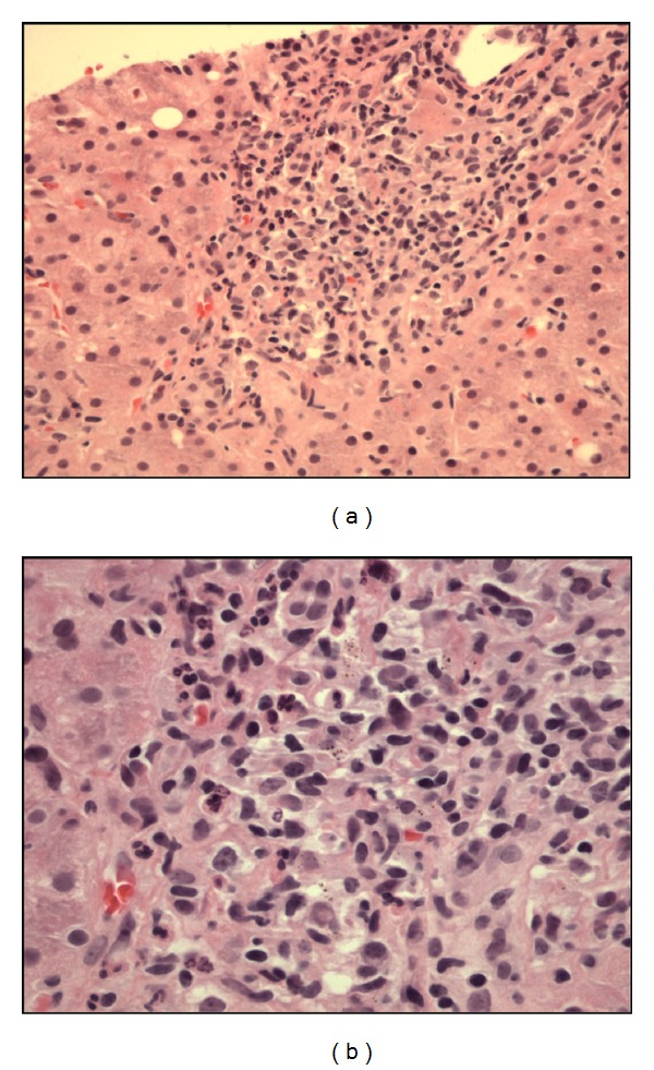
(a) Liver biopsy specimen on H&E stain showing noncaseating granuloma. (b) On greater magnification, the granuloma consists of mixture of inflammatory cells, including epithelioid histiocytes and lymphocytes.

(a) Liver biopsy specimen on H&E stain showing noncaseating granuloma. (b) On greater magnification, the granuloma consists of mixture of inflammatory cells, including epithelioid histiocytes and lymphocytes.