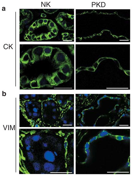
Figure 2. Vimentin is anomalously expressed in cystic epithelia in situ.
a. Cryosection of normal kidney showing cytokeratin (CK) staining in tubule cells (NK) Lower panel is a higher magnification of upper one. ADPKD kidney sectioned at a cyst level (PKD) shows that cells lining the wall cyst conserve cytokeratin expression (magnified in lower panel). Samples were imaged on Zeiss LSM510. Bars, 20 μm.
b. Cryosection of normal tubules (NK) labeled against vimentin protein (VIM), show complete a absence of vimentin from their cytoplasm (magnified in lower panel). Kidney interstitial fibroblasts have a normal vimentin signal. Sections from a cyst of an ADPKD kidney (PKD) show the abnormal presence of vimentin in cyst-lining epithelial cells (detailed in lower panel). Samples were imaged on Zeiss LSM510. Bars, 20 μm.

