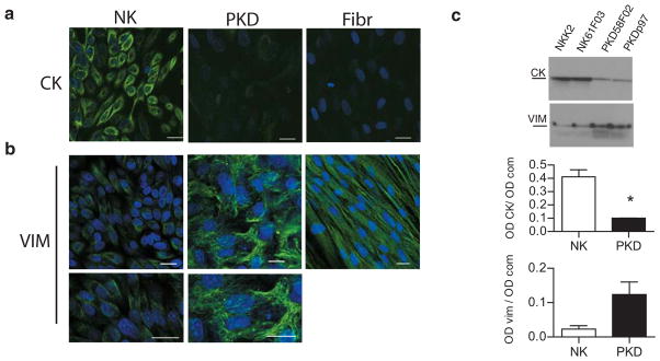
Figure 3. ADPKD cells express an immature intermediate filament network even when fully confluent.
a. Confocal images of primary normal kidney cells (NK) and polycystic kidney cells (PKD) in culture labeled against cytokeratin intermediate filament (CK). Normal kidney cells show normal cytokeratin labeling, while disease cells show very low signal. Confocal images of normal and polycystic kidney cells in culture, labeled against vimentin intermediate filament (VIM). Normal cells have very little vimentin signal (magnified in bottom panel), but polycystic cells, on the contrary, show a conspicuous vimentin filament network (amplified in lower panel). Normal human fibroblasts (Fibr) express exclusively vimentin. Bars, 20 μm.
b. Immunoblots of lysates from two different samples each of normal (NK) and ADPKD (PKD) kidney cells probed for cytokeratin (CK) or vimentin (VIM). Below, quantification of cytokeratin and vimentin protein expression levels in normal and disease cells grown. The y-axis represents the intensity of the CK or the VIM band (OD CK) normalized according to the total protein loaded per lane as detected by Coomassie blue staining (OD com). CK graph: p=0.01. Vim graph: p=0.057.

