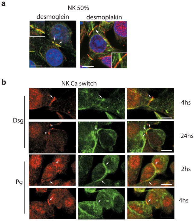
Figure 6. Polycystin-1 and desmosomal proteins transiently colocalize when normal kidney cells are at 50% confluence.
Confocal images of normal primary human kidney cells taken at early stage of cell-cell contact. Co-labeling of polycystin-1 (red) and desmosomal components (green) was carried out.
a. Cells processed at 50% confluence show an overlapping pattern (arrows) between polycystin-1 and desmoglein (left panel) or desmoplakin (right panel). Insets show a magnification of membrane region marked with arrows. Bars, 10 μm.
b. Cells were subjected to a calcium switch assay and processed at different time points after the calcium switch. Desmoglein staining (upper two rows) shows clear colocalization with polycystin-1 at 4 h following the calcium switch. This colocalization is lost after 24 h. Plakoglobin labeling (lower two rows) shows colocalization with polycystin-1 at 2 and 4 h after the switch to normal calcium. Bars, 10 μm.

