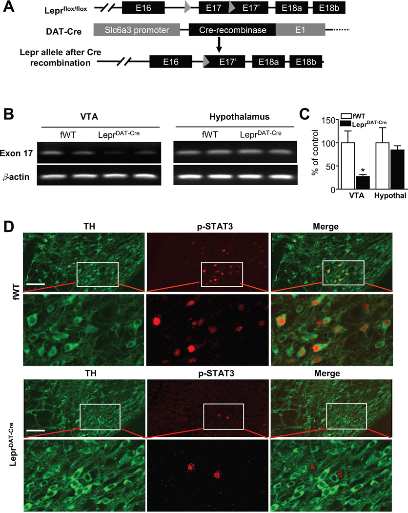
Figure 1.
Generation of mice lacking Lepr selectively in dopamine neurons. (A) Schematic diagram depicting the floxed Lepr allele, the Slc6a3 (or DAT) Cre allele, and the Lepr floxed allele after recombination. (B) RT-PCR detection of exon 17 of the leptin receptor in the ventral tegmental area (VTA) versus hypothalamus in LeprDAT-Cre mice and Leprflox/flox littermate control (fWT) mice. (C) Real-time quantitative PCR analysis showing a Cre-mediated deletion of exon 17 of Lepr in the VTA of LeprDAT-Cre mice. Values are expressed as a percentage change from fWT control mice. Data are expressed as mean ± SEM. n = 4 per group. *p < 0.05 compared with fWT control mice. (D) Double-labeling immunohistochemistry showing the colocalization of phosphorylated STAT3 in dopamine neurons, positive for tyrosine hydroxylase (TH), in fWT control mice and LeprDAT-Cre mice. The loss of Lepr in dopamine neurons eliminates leptin-stimulated phosphorylation of STAT3 in dopamine neurons in LeprDAT-Cre mice. Scale bar = 100 µM.

