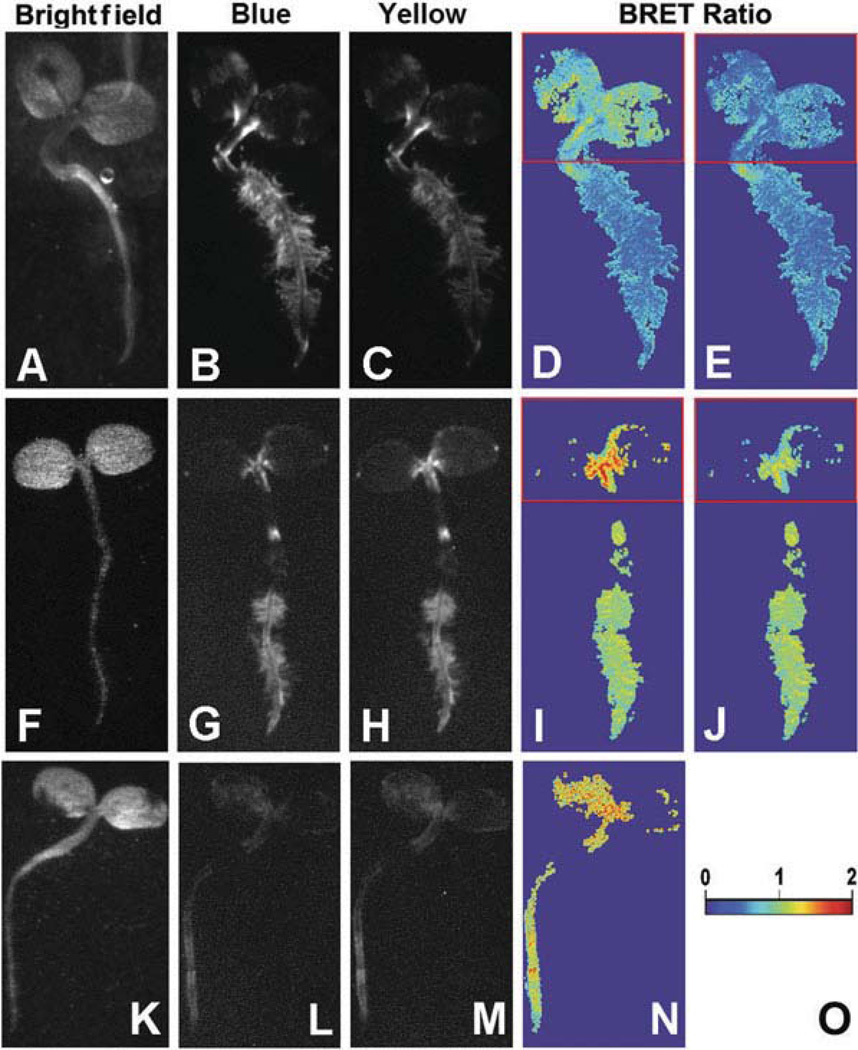
Fig. 1.4. BRET macro-imaging of tobacco seedlings.
Seven-day-old tobacco seedlings were transformed with (i) P35 s::Rluc (a–e), (ii) P35s::Rluc-EYFP (f–j), or (iii) P35 s::Rluc-COP1+ P35 s::Eyfp-COP1 (k–n). Panels a, f, k are bright field images, panels b, g, l are images of short-pass luminescence (Blue), panels c, h, m are images of long-pass luminescence (Yellow), panels d, i, n are BRET ratios (Y ÷ B) over the entire luminescent portion of the image (pseudocolor scale shown in panel o), panels e and j are corrected images of panel d and i, respectively, shown with a red box encasing the pigmented (cotyledon) portion of the seedlings (correction factor for boxed regions of panels e and j was 1.27). (Modified from Xu et al. (16).)

