
Figure 4.
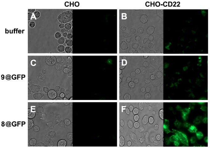
Phase contrast and fluorescent microscope images of CHO (A, C and E) and CHO-CD22+ cells (B, D and F) incubated with PBS (A and B), 9@GFP16 (C and D), and 8@GFP16 (E and F) at 37 °C for 4 hours. Each particle was used at 1 nM (50 nM in porphyrin); the @ symbol denotes the encapsidation of multiple copies of GFP inside the particle. The faint signals observed in panels A and B are due to cellular autofluorescence.