
Figure 6.
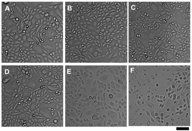
Phase contrast microscopy of CHO-CD22 cells after treatment with particles and light as described in Figure 5. (A-C) Cells incubated with LacNAc-loaded Qβ particles 9. (D-F) Cells incubated with BPC ligand-loaded Qβ particles 8. (A,D) Cells incubated with 10 nM particle for 4 h at 37°C. (B,E) After incubation with particle, washing, and irradiation (430 ± 10 nm, 90 min, RT). (C,F) After incubation with particle, washing, irradiation, incubation in CO2 incubator overnight at 37°C. Scale bar = 50 μm.