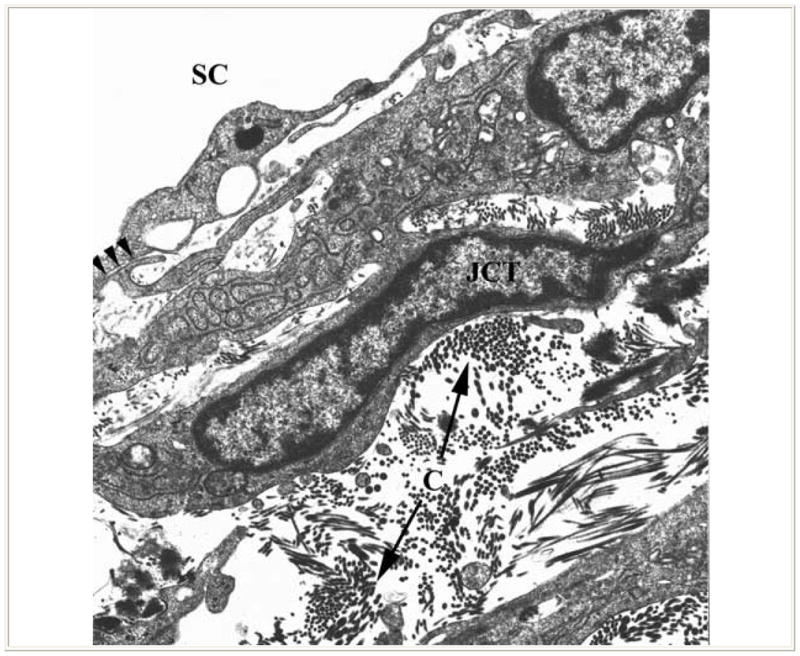
Figure 2.

Transmission electron micrograph shows juxtacanlicular region (JCT) of the trabecular meshwork and inner wall of Schlemm’s canal (SC). The JCT region exhibits an open matrix including collagen (C) and elastin. The fibroblast-like cells of the region extend slender connections to the endothelial cells lining Schlemm’s canal (arrows). (From Freddo T. Chapter 3. Ocular anatomy and physiology related to aqueous production and outflow. In: Primary Care of the Glaucomas. Lewis T, Fingeret M, eds, Appleton and Lange, 1993. With kind permission of The McGraw-Hill Companies Inc.)
