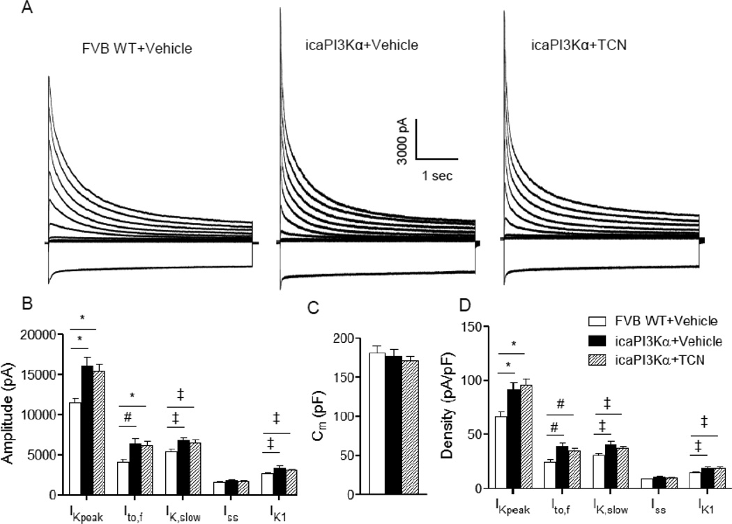
Figure 6. Short-term activation of cardiac PI3Kα signaling upregulates repolarizing K+ currents in an Akt-independent manner.
(A) Representative whole-cell K+ currents, recorded from LV apex myocytes isolated from WT+Vehicle, icaPI3Kα+Vehicle and icaPI3Kα+TCN animals, are shown. (B) Mean ± SEM IK,peak, Ito,f, IK,slow and IK1 amplitudes in icaPI3Kα+Vehicle (n=24) were markedly higher than in WT+Vehicle (n=33) LV apex myocytes; K+ current (IK,peak, Ito,f, IK,slow and IK1) amplitudes were also significantly higher in icaPI3Kα LV apex myocytes treated with TCN (n=21). (C) Mean ± SEM Cm values were similar in WT+Vehicle, icaPI3Kα+Vehicle and icaPI3Kα+TCN LV apex myocytes, consistent with the absence of cellular (LV) hypertrophy with short term caPI3Kα induction (see text). (D) Normalization of current amplitudes for differences in cell size (Cm) revealed that mean ± SEM IK,peak, Ito,f, IK,slow and IK1 densities were significantly (‡P<0.05, #P<0.01, *P<0.001) higher in icaPI3Kα+vehicle, than in WT+Vehicle, LV apex myocytes. The higher K+ current densities observed in icaPI3Kα LV apex myocytes were unaffected by the TCN treatment.

