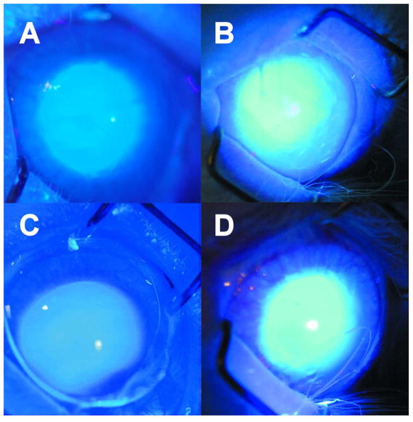
Figure 1.
Photography taken during exposure of a rabbit eye to UVA radiation in vivo (365 nm, 100 mW/cm2 on the cornea). A. No contact lens; after 1 min of irradiation, B. No contact lens; after 1 hour of irradiation, C. senofilcon A lens; 1 hour of irradiation with the contact lens, followed by removal of the contact lens, continuation of irradiation for 1 min and photography, D. lotrafilcon A lens; 1 hour of irradiation with the contact lens, followed by removal of the contact lens, continuation of irradiation for 1 min and photography. Representative of 3–6 experiments.

