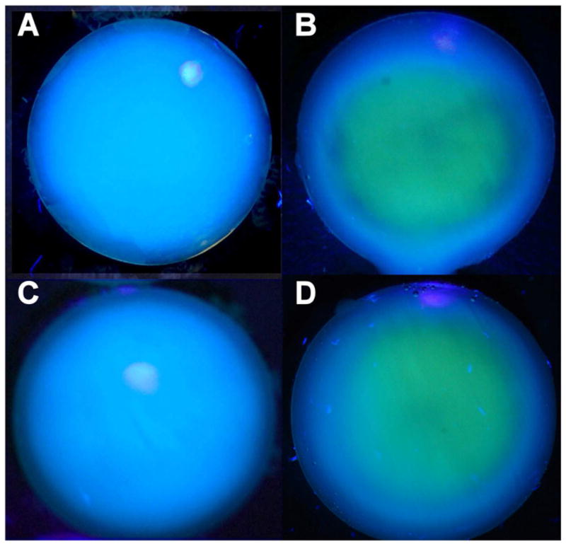
Figure 2.
Photography taken during exposure of isolated rabbit lenses of Figure 1 to UVA light (365 nm, 100 mW/cm2). A. Normal lens, B. Prior exposure for 1 hr in vivo with no contact lens (Fig. 1B), C. Prior exposure for 1 hr in vivo with a senofilcon A lens (Fig. 1C), D. Prior exposure for 1 hr in vivo with a lotrafilcon A lens (Fig. 1D). Representative of 3–6 experiments.

