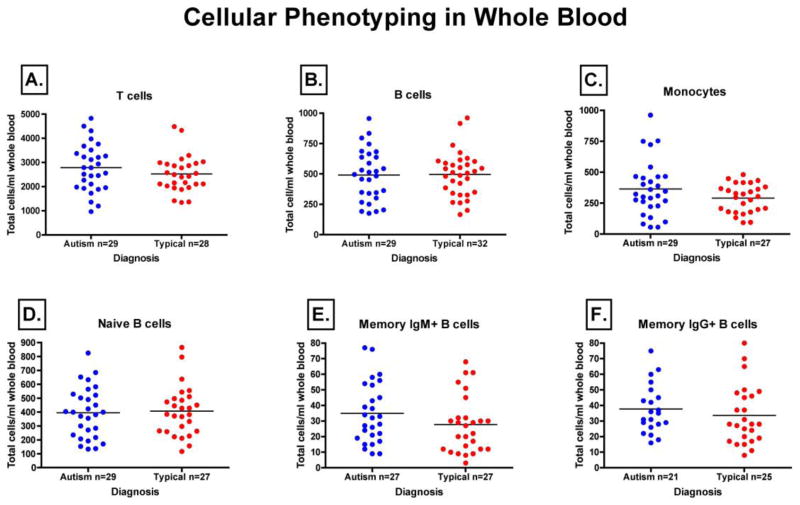
Figure 3. Median quantities of immune cell subsets in peripheral blood as determined by flow cytometry.
Whole blood from both autism and typically developing controls were stained using fluorochrome-conjugated antibodies directed against cell surface phenotypic markers. Absolute quantification of peripheral blood immune cell populations was then carried out by flow cytometry using counting beads. We found no significant differences in the absolute quantity of CD19+ B cells (A), CD3+ T cells (B), CD14+ Monocytes (C), CD19+CD27−IgD+ Naïve B cells (D), CD19+CD27+IgM+ Memory B cells (E), or CD19+CD27+IgG+ Memory B cells (F).

