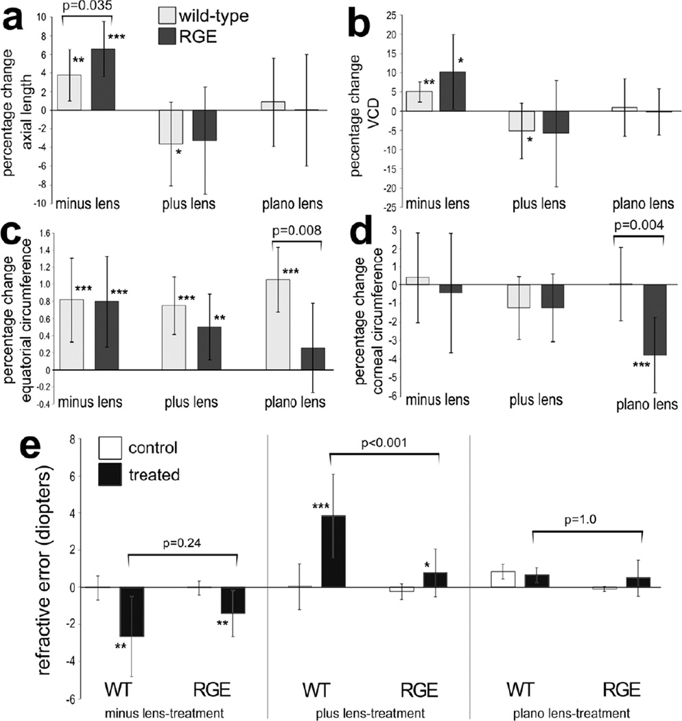
Figure 2.
Lens-induced changes in ocular growth in WT and RGE eyes. The right eyes of WT and RGE chickens were treated with minus 7 lenses (n=10), plus 7 lenses (n=10) or plano lenses (n=6) for 4.5 days beginning at P7, and left eyes were used as controls. Measurements of axial length (a) and vitreous chamber depth (b) were made using A-scan ultrasound. Measurements of equatorial circumference (c) and corneal circumference (d) were obtained by analysis of digital images with ImagePro 6.2. The mean percentage change between treated and control ocular parameters were calculated. Retinoscopy was used to determine refractive error in WT and RGE eyes treated minus lens-wear, plus lens-wear or plano lens-wear. Error bars represent standard deviation. Significance of difference (*p<0.05, **p<0.01, ***p<0.001) for percentage change of control vs treated within a group was determined by using a Student’s t-test. Significance of difference between treatment groups was determined by using a two-way ANOVA (p<0.001) and a post-hoc Bonferroni analysis. Asterisks indicate significant differences within a treatment group (percent change), and brackets and p-values indicate significant differences between treatment groups (WT vs RGE). Abbreviations: WT – wild type, RGE – retinopathy globe enlargement, VCD – vitreous chamber depth.

