Fig. 1.
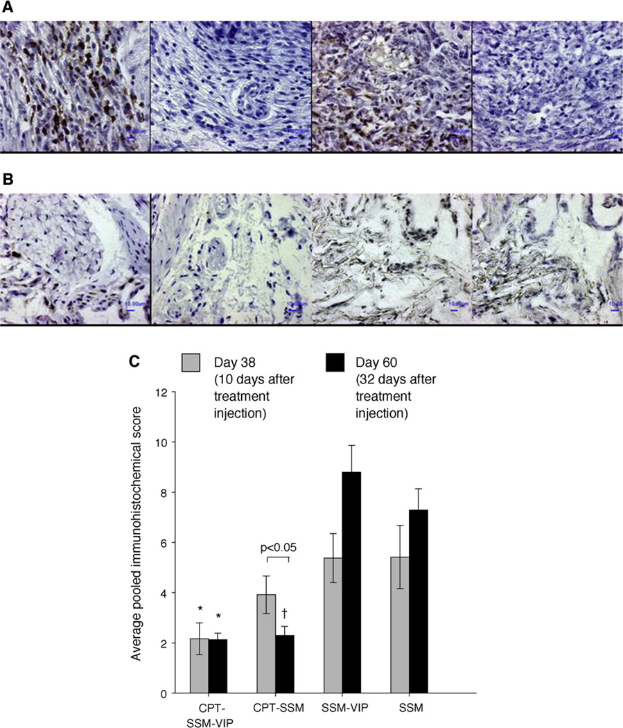
CPT in micelles reduces cellular content within the CIA joints. Representative joint sections of (A) empty SSM-VIP-injected mice, (B) CPT-SSM-VIP treated mice; (left to right) CD3+, CD3−, lysozyme+, lysozyme− staining. Bars represent 10 µm. (C) Average pooled immunohistochemical scores of cellular infiltration and distribution in joints of CIA mice on ( ) Day 38 and (■) Day 60. Results are expressed as mean ± S.E.M. (6 mice/group). *, †p< 0.05 versus SSM-VIP and SSM, respectively.

