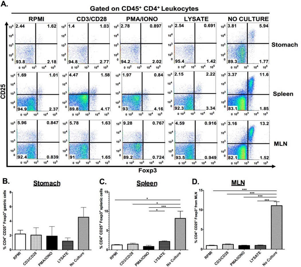
Figure 3. Effect of ex vivo stimulation on detection of regulatory T cells.
Lymphocytes isolated from the gastric, splenic and mesenteric lymph node (MLN) compartments were cultured under either indirect (CD3/CD28, PMA/IONO) or direct stimulation (H. pylori lysate) and immunophenotype for CD25+ Foxp3+ regulatory T cells. Representative dot plots demonstrated that ex vivo stimulation yielded a decrease in the percentage of regulatory T cells regardless of the compartment the cells originated from. (A) This observation was quantified, and indicated that ex vivo stimulation significantly abrogates detection of CD4+ CD25+ Foxp3+ regulatory T-cells (B–D). Graphs represent the mean ± standard error of 4 mice pooled per experiment; results representative of 3 independent experiments. Groups were compared using one-way analysis of variance (ANOVA) followed post-hoc by Tukey multiple comparisons tests on log-transformed data. Significant differences are represented by the following *p<0.05, **p<0.01, ***p<0.001.

