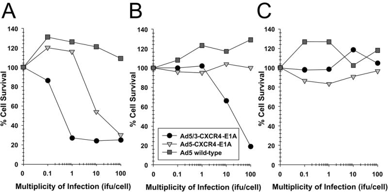
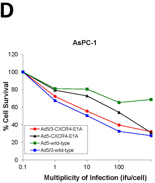
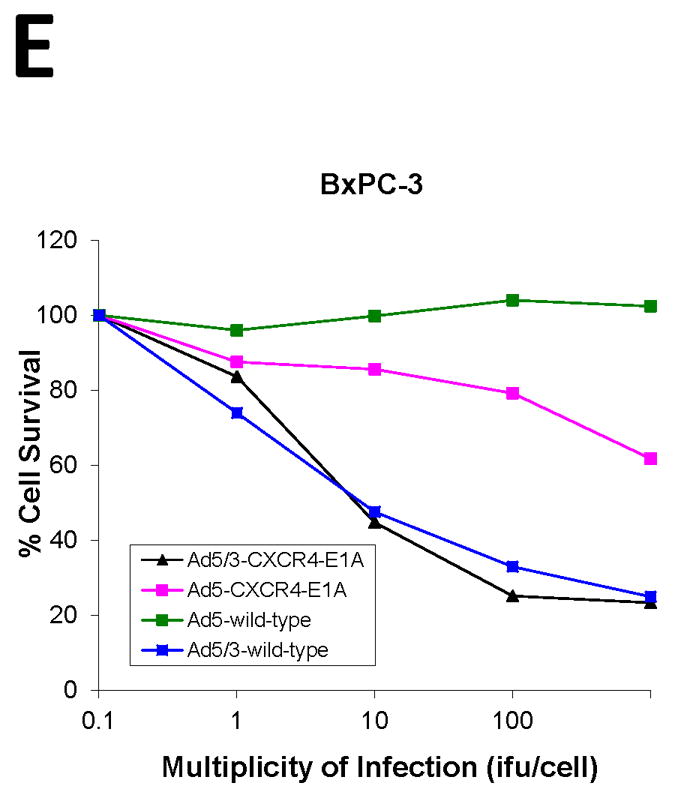
FIGURE 3. Oncolytic activity of an oncolytic Ad construct in pancreatic cancer cell lines.


Oncolysis was evaluated by crystal violet staining in the pancreatic cancer cell lines (A) PANC-1, (B) CFPAC-1, (C) MAT BIII, (D) AsPC-1, (E) BxPC-3 after infection with increasing doses from 1 i.fu./cell to 100 i.fu./cell of Ad5/3-CXCR4-E1A (●), Ad5-CXCR4-E1A (▼), or Ad5 wild-type (■) as described in Material and Methods. Values represent the mean ± SEM of 3 replicate measurements.
