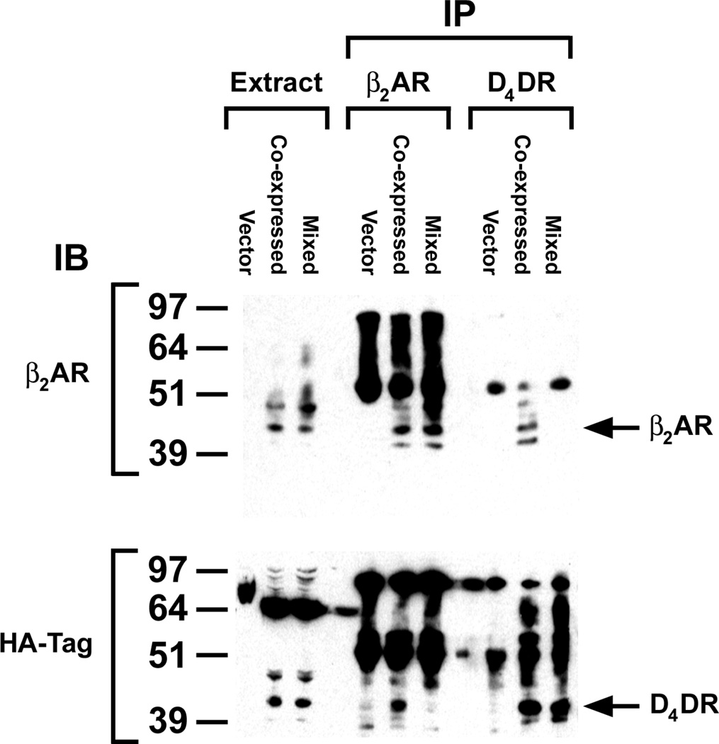
Figure 1. Co-immunoprecipitation of exogenously expressed β2AR and D4DR.
All cells used in these studies transiently co-expressed Gαs, Gαi1, Gβ1, Gγ2 and AC. In addition they expressed β2AR and/or the HA-tagged D4DR. Cells expressing one or the other receptor were mixed prior to preparing membranes (Mixed). So that each sample contained the same ratio of expressed receptor protein to total cell protein, cells co-expressing the receptors (Co-expressed) were mixed with cells receiving plasmid without receptor cDNA. These latter cells (Vector) were also used as a negative control. Membranes prepared from these samples were dissolved in RIPA buffer, and the soluble proteins in the supernatant following centrifugation at 100,000 × g were treated as described in Materials and Methods in order to immunoprecipitate and identify β2AR and HA-tagged D4DR on western blots. Data are representative of three independent experiments.

