
Figure 1.
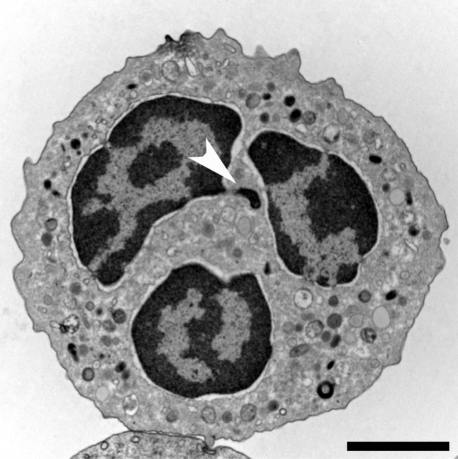
Neutrophil morphology. Transmission electron microscopy (TEM) of a naive human neutrophil. This cell contains various types of granules, clearly visible in the cytoplasm, as well as a lobulated nucleus. The highly condensed heterochromatin (dark) is neatly marginalized to the edge of the nucleus, only interrupted by euchromatic areas close to nuclear pores that mostly line the nuclear membrane. The brighter euchromatin is mostly in the center of the lobules. This neutrophil comes from a female donor and one inactivated x chromosome can be found as an extranuclear stretch of heterochromatin (arrowhead). These structures are termed Barr bodies, and in neutrophils “drum sticks.” Bar, 2 µm.