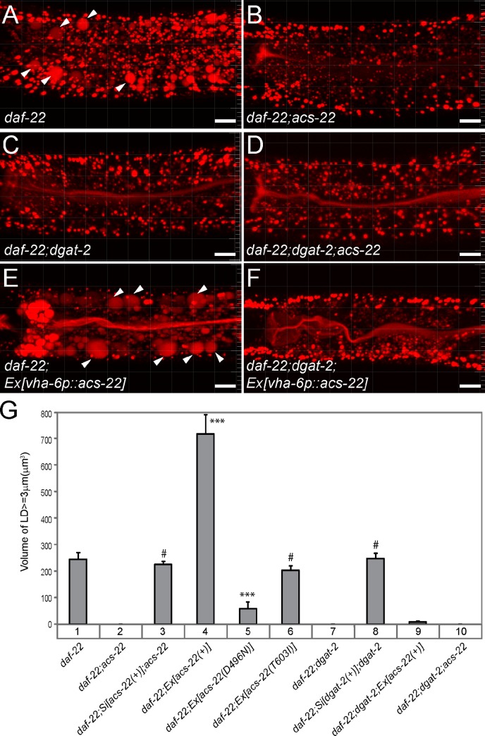
Figure 2.
ACS-22/FATP1 and DGAT-2 act in the same genetic pathway. (A–F) Red BODIPY-C12 staining of larval stage L4 animals. Expanded LDs >3 µm in diameter were indicated by white arrowheads. Images were 3D projections of 9-µm confocal z stacks that covered the first three intestinal segments. Bars, 10 µm. (G) Quantification of total volumes of BODIPY-positive structures that were >3 µm in diameter in the second intestinal segment of larval stage L4 animals. For each strain, a total of 20 animals were imaged at two independent times, except for daf-22; hjSi29[acs-22(+)]; acs-22 (n = 17) and daf-22; hjSi56[dgat-2(+)]; dgat-2 (n = 16). For experiments using extrachromosomal arrays, two independent transgenic lines were quantified, with 10 animals each. Data were plotted as means ± SEM. Pairwise t test between lane 1 and lanes 3, 6, or 8: #, P > 0.1. Pairwise t test between lane 1 and lanes 2, 4, 5, 7, 9, or 10: ***, P < 0.001.

