Figure 3.
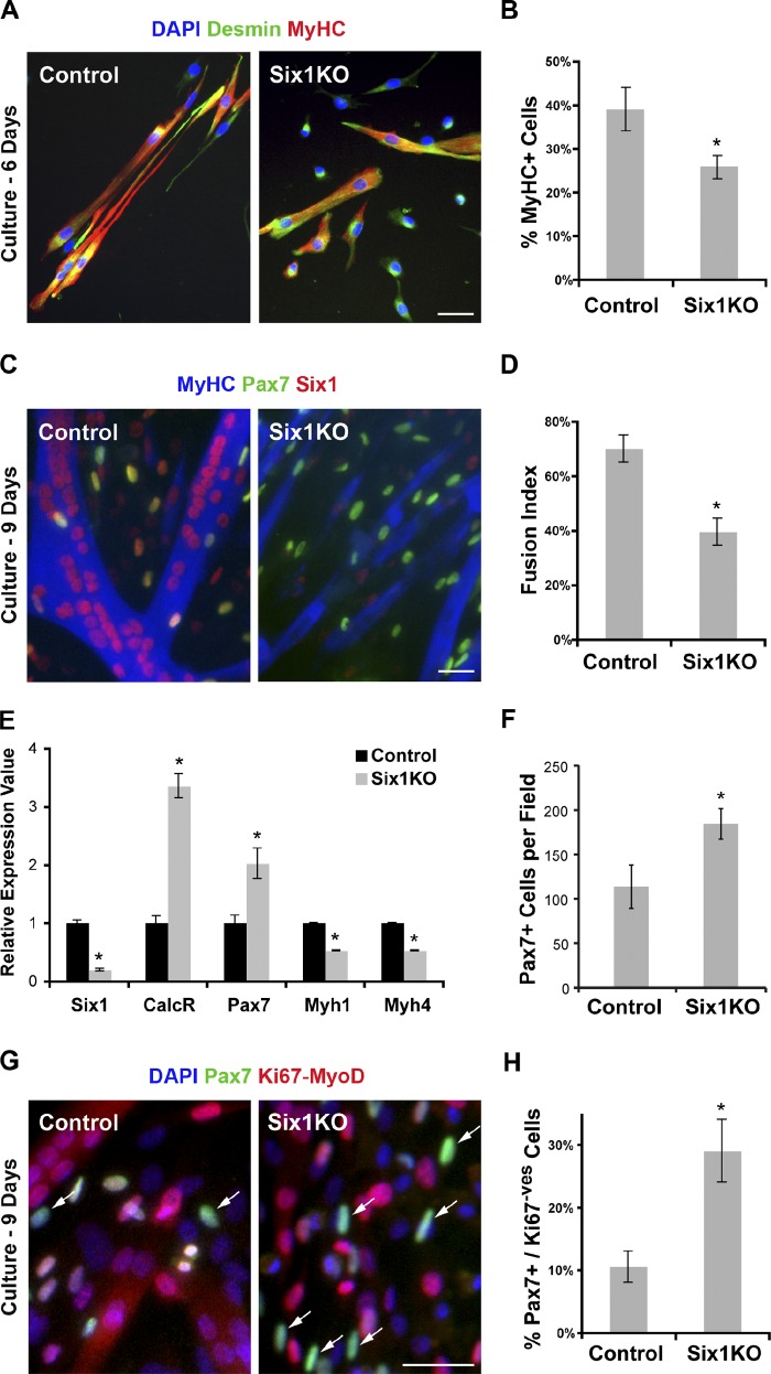
Six1 gene disruption perturbs myogenic differentiation of SC descendants ex vivo. 1 wk after TM treatment, EDL myofibers from control and Six1KO mice were plated on Matrigel, and cultures were analyzed after 6 and 9 d of culture ex vivo. (A) Myogenic cells grown for 6 d were immunolocalized for Desmin (myoblast marker) and MyHC (differentiation marker) proteins. (B) Six1KO cells exhibit limited differentiation potential ex vivo compared with control cells. (C) Myogenic cells grown for 9 d were immunolocalized for Six1, Pax7 (undifferentiated state marker), and MyHC (differentiated state marker) proteins. (D) Six1KO cells fuse less efficiently and form smaller myotubes compared with control cells. (E) qRT-PCR analysis indicated expression of Six1, CalcR, Pax7 (SC markers), and Myh1, Myh4 (differentiation markers) transcripts by differentiated myogenic cells. (F) Six1KO cell cultures generate more Pax7+ cells compared with control cells. (G) SC-derived myogenic cells grown for 9 d were immunolocalized for Pax7 and both MyoD and Ki67 proteins. (H) Six1KO cells generate more “reserve” cells (Pax7+/MyoD−/Ki67−; arrows) compared with control cells. Error bars indicate standard deviations. *, P < 0.02. Bars, 10 µm.

