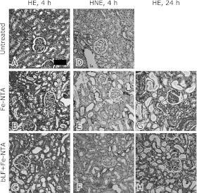
Fig. 4.
Histological and immunohistochemical analyses of kidney at 4 and 24 h after Fe-NTA administration. Hematoxylin and eosin staining (HE). (A) Untreated, (B) Fe-NTA alone, 4 h, (C) or in the presence of bLF + Fe-NTA, 4 h. Immunohistochemical staining of 4-hydroxy-2-nonenal-modified proteins (HNE). (D) Untreated, (E) Fe-NTA alone, 4 h, (F) bLF + Fe-NTA, 4 h. HE staining. (G) Fe-NTA alone, 24 h, and (H) bLF + Fe-NTA, 24 h. Representative images are shown. Scattered necrotic tubules were detected (B). Only a few necrotic tubules and some degenerative tubules were observed (C). HNE immunostaining revealed accumulation of oxidatively modified proteins. No positive tubules in the HNE immunostaining were observed (D), whereas many positive tubules were detected (E). Immunopositivity was markedly decreased (F). After Fe-NTA administration for 24 h, oxidative injury destroyed massive proximal tubules (G). Pretreatment with bLF protected against oxidative injury (H). Few infiltrating inflammatory cells were observed in histological samples (bar, 50 µm).

