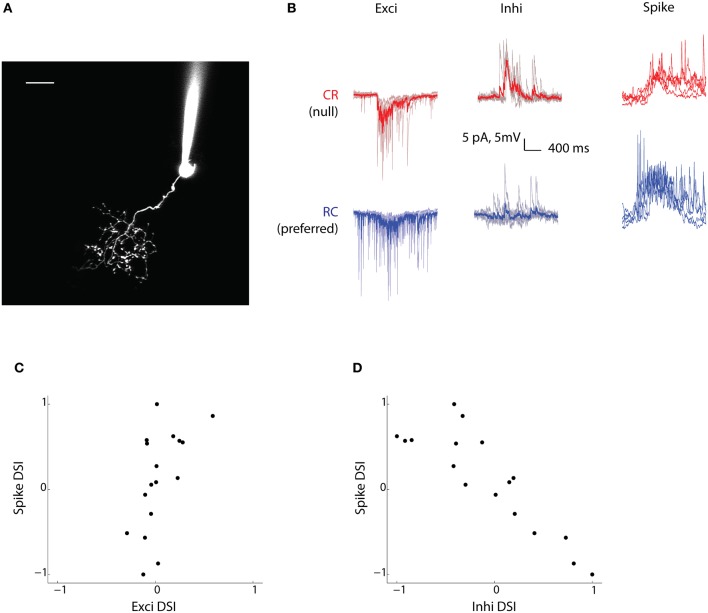
Figure 3.
Inhibitory currents are biased toward the null direction of motion. (A) Two-photon fluorescence image of a tectal neuron filled with dye (Alexa 594) from the recording electrode. Tectal neurons project dendritic arbors into a neuropil where they receive retinal ganglion cell axonal input from the contralateral eye. Scale bar represents 10 μm. (B) Voltage clamp (Exci and Inhi) and current clamp (Spike) recordings of a tectal cell's response (for the duration the stimulus was presented) to bars moving in the CR direction (red) and the RC direction (blue). For the currents, the mean trace (thicker red/blue line) is shown superimposed over five trials (lighter red/blue lines). This cell is RC selective and has strong inhibition from the null CR direction. (C and D) Spike-DSI for all cells (n = 17) plotted vs Exci-DSI and Inhi-DSI, respectively.

