
Figure 8.
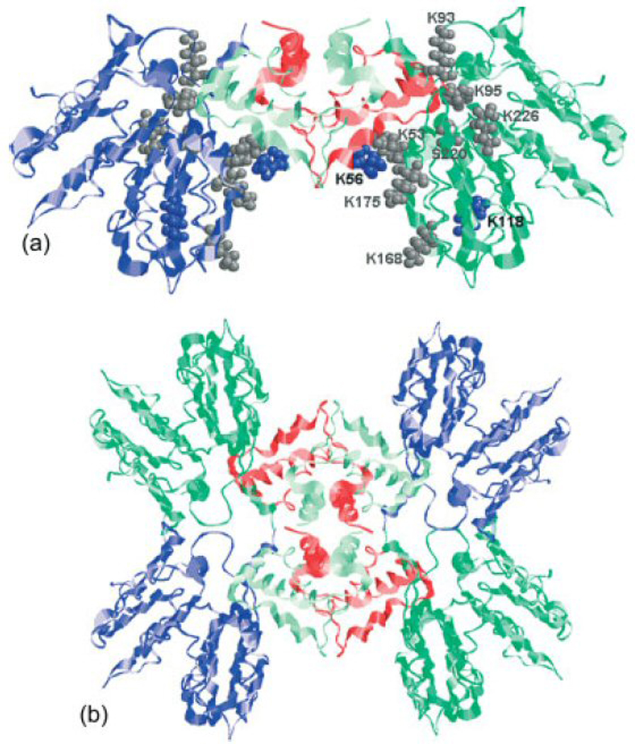
(a) MS3D model of the ~100 kDa ANXA2/P11 complex.222 The location of lysine residues involved in intra- and inter-protein crosslinking of the p11 dimer and full-length ANXA2 are marked as blue and grey spheres. (b) These crosslinks were used for docking calculations that provided a hetero-octameric A2t complex consisting of four ANXA2 (dark green and blue) located on the periphery connected by two p11 dimers (red and light green) in the center. Adapted with permission from reference 222.