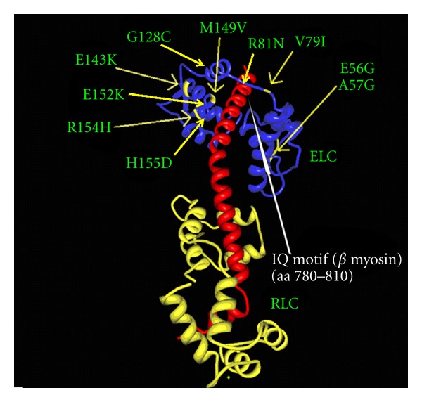
Figure 5.

The location of the p.V79I mutation (arrow) in the three-dimensional structure of the regulatory domain of myosin (red) with the essential (ELC) (blue) and regulatory (RLC) (yellow) light chain. The other known mutations in ELC are also marked. It is seen that the p.V79I mutation; as well as the pR81H and p.M149V mutation, is located close to the IQ1 motif of the myosin helix. The IQ1 motif is marked by a white arrow. The figure is based on X-ray crystallographic structure of the myosin myosin regulatory domain of the scallop as given in the 1WDC pdb file [32].
