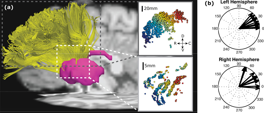
Figure 1.
Fiber tracts between dorsolateral PFC and striatum from human diffusion tractography. From [10]. (a) Fibers in an example subject that start in the dorsolateral PFC (top inset) and terminate in the striatum (bottom inset) as seen in the sagittal plane (D, dorsal; V, ventral; R, rostral; C, caudal). Color gradient shows start from more rostral (blue) to caudal (red) along the frontal gyrus. (b) Vectors show shifts in fiber position in the striatum as cortical start position goes rostral to caudal in the sagittal plane for ten subjects.

