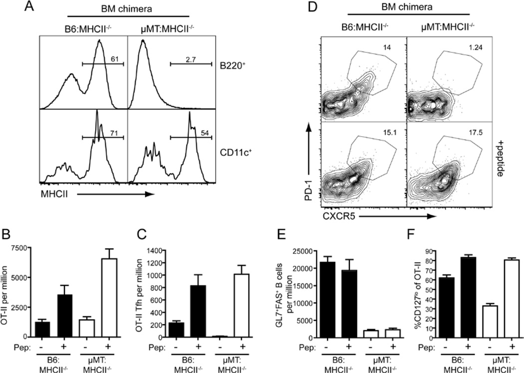
Figure 5. B cell Ag-presentation is not required for Tfh development.
Chimeras were generated as described in Experimental Procedures using 80:20 mix of µMT:MHCII−/− BM (to generate mice with B cells lacking MHC class II) or B6:MHCII−/− BM (controls). Thy1.1+ WT OT-II cells were transferred into the chimeras, which were then given OVA plus Alum i.p. at day 0. On day 3 some mice received additional OVA peptide i.v. and the mice were sacrificed on day 7 for analysis. (A) Reconstitution was determined by staining B cells (B220+) and DC (CD11c+) for MHCII expression. (B) The proportion of total OT-II cells in the spleen. (C) The proportion of CXCR5hiPD1hi Tfh OT-II cells was determined by (D) staining for CXCR5 and PD1 on OT-II cells. (E) Proportion of GC B cells as assessed by staining for expression of GL7 and Fas. (F) Percentage of cells OT-II cells that are CD127lo. All plots show Mean ± SEM, n=6–8.

