
Figure 4.
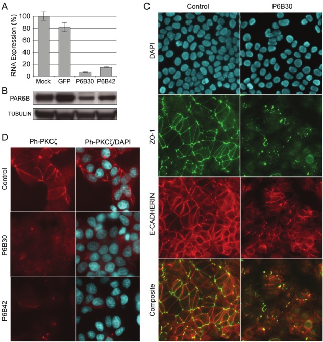
Inhibition of PAR6B in MCF7 cells disrupts tight junction but not adherens junction assembly. A. Quantitative RTPCR analysis of PARD6B expression level in MCF7 cells following siRNA electroporation and 24h incubation. siRNAs were control, and PARD6B-spedcific P6B30 and P6B42. Mock indicates electroporated cells without siRNA. B. Western blot to detect PAR6B protein in MCF7 whole cell lysate following 24h siRNA treatment. Immunoblot was re-probed with anti-tubulin antibody to control for protein loading. C. Dual immunofluoresence stain of MCF7 cells for ZO-1 and E-cadherin following siRNA electroporaiton and 24h incubation. SiRNAs are control or PARD6B-spedcific P6B30. Composite images show the merge of ZO-1 and ECAD stains without DAPI. AJ networks are intact following PAR6B knockdown, whereas TJ networks are lost. D. IF stain for phospho-PKCζ in MCF7 cells following siRNA-mediated knockdown of PAR6B.