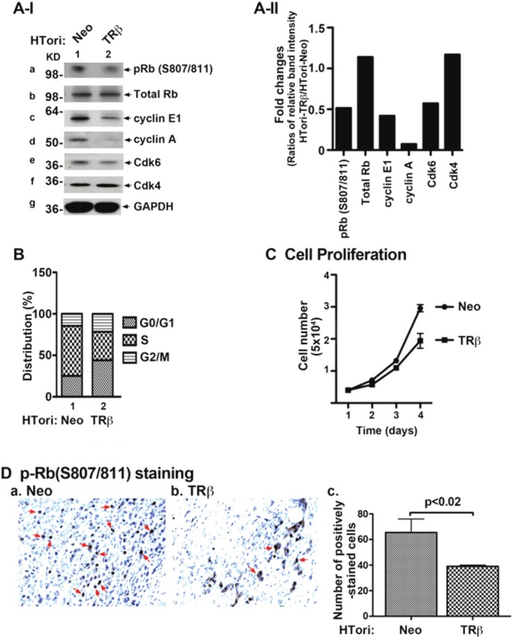
Figure 4.

Reactivation of Rb by physical interaction of TRβ with SV40Tag. (A-I) HTori-Neo and HTori-TRβ cells were grown in 60mm dish for 24 hours and the cell extracts were prepared, followed by Western blot analyses (20 μg cell lysates were used) for key regulators of cell cycle progression (as marked), as described in Materials and Methods. The reactivation of Rb was indicated by decreased phosphorylation (panel a), cyclin E1 (panel c), cyclin A (panel d), and Cdk 6 (panel e). (A-II) Fold of changes in the protein abundance of key regulators in HTori-TRβ cells as compared with those in HTori-Neo cells. Each protein level was normalized to GAPDH. (B) Cell cycle distribution was determined in HTori-Neo cells (bars 1) and HTori-TRβ cells (bars 2), as described Materials and Methods. Delayed entries of cells from the G1 to the S phase were observed in cells expressing TRβ. (C) Proliferation of HTori-TRβ cells was less than that of HTori-Neo cells. Data are the mean ± SEM; n=3. (D) pRb (S807/811) staining of tumor cells derived from HTori-Neo cells (panel a) and cells derived from HTori-TRβ (panel b). Arrows point to the positive staining of pRb (S807/811). The positively pRb (S807/811) stained cells were counted and graphed (panel c). The difference in the number of positively stained cells is highly significant (p<0.02).
