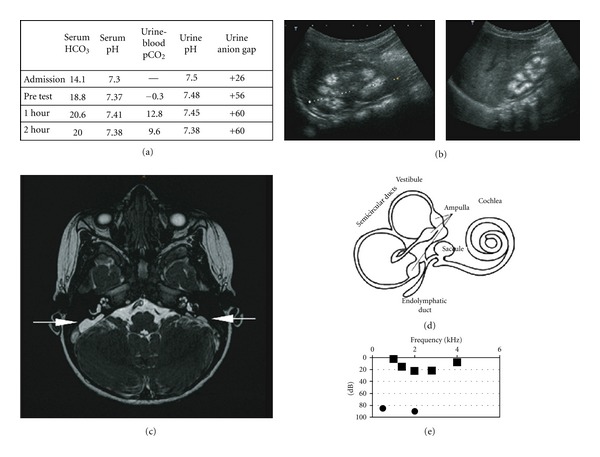
Figure 1.

(a) Results of bicarbonate loading test with 1 mmol/kg NaHCO3 infusion. (b) Ultrasound of the left and right kidney with evidence of nephrocalcinosis. (c) Axial FIESTA cerebral MRI: bilateral enlargement of the endolymphatic sac (arrows), markedly larger on the right. (d) Schematic image of the inner ear. (e) Results of free field measurement and inserted right ear audiogram.
