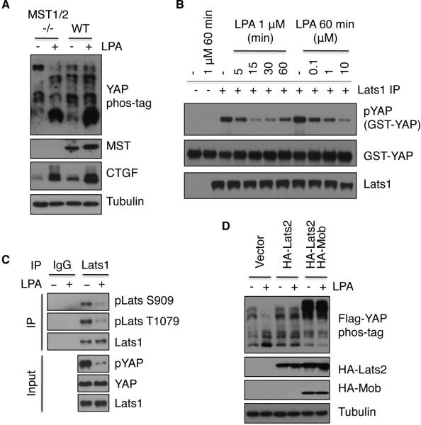
Figure 5. LPA and S1P repress Lats kinase activity.
(A) MST1/2 are not required for LPA-induced YAP dephosphorylation and CTGF induction in MEF cells. WT or knockout MEF cells at similar density were untreated or treated with 1 μM LPA for 1 h. YAP phosphorylation was assessed by immunoblotting in the presence of phos-tag. (B) Lats kinase activity is inhibited by LPA. Endogenous Lats1 was immunoprecipitated from HEK293A cells that had been treated with LPA at various times and doses of LPA, and Lats1 kinase activity was determined using GTS-YAP as a substrate. (C) Lats phosphorylation is repressed by LPA. Cell lysates from control or LPA-treated (1 μM for 1 h) cells were divided into two parts, one for IgG IP and the other for Lats1 IP. Endogenous Lats1 was immunoprecipitated and probed with phospho-specific antibodies. (D) Lats overexpression suppresses the effect of LPA on YAP phosphorylation. HEK293A cells were co-transfected with Flag-YAP and HA-Lats2 or HA-Mob. One day after transfection, cells were serum-starved for 24 h, and then treated with 1 μM LPA for 1 h. Also see also Figure S5.

