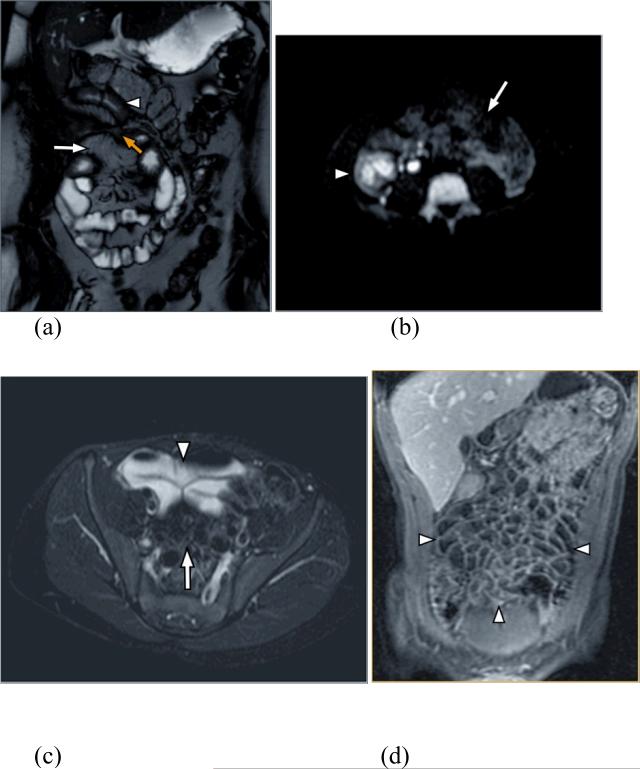
Figure 1.
(a) Coronal FIESTA allows depiction of bowel wall thickening (arrowhead) and the mesenteric fat and vessels (log arrow). Yellow arrow depicts enteroenteric fistula. (b) Diffusion weighted images showing restricted diffusion due to edema in the right lower quadrant (arrowhead) in contrast to normal bowel with no restricted diffusion in the left side of the abdomen (log arrow). (c) Axial T2 weighted image shows high signal of the intraluminal fluid within the lumen (arrowhead) with good depiction of the mesenteric fat (log arrow). (d) Coronal T1 weighted 3D SPGR after contrast administration shows the contrast between the hypointense lumen and the enhancing bowel wall improving the ability to assess abnormal enhancement

