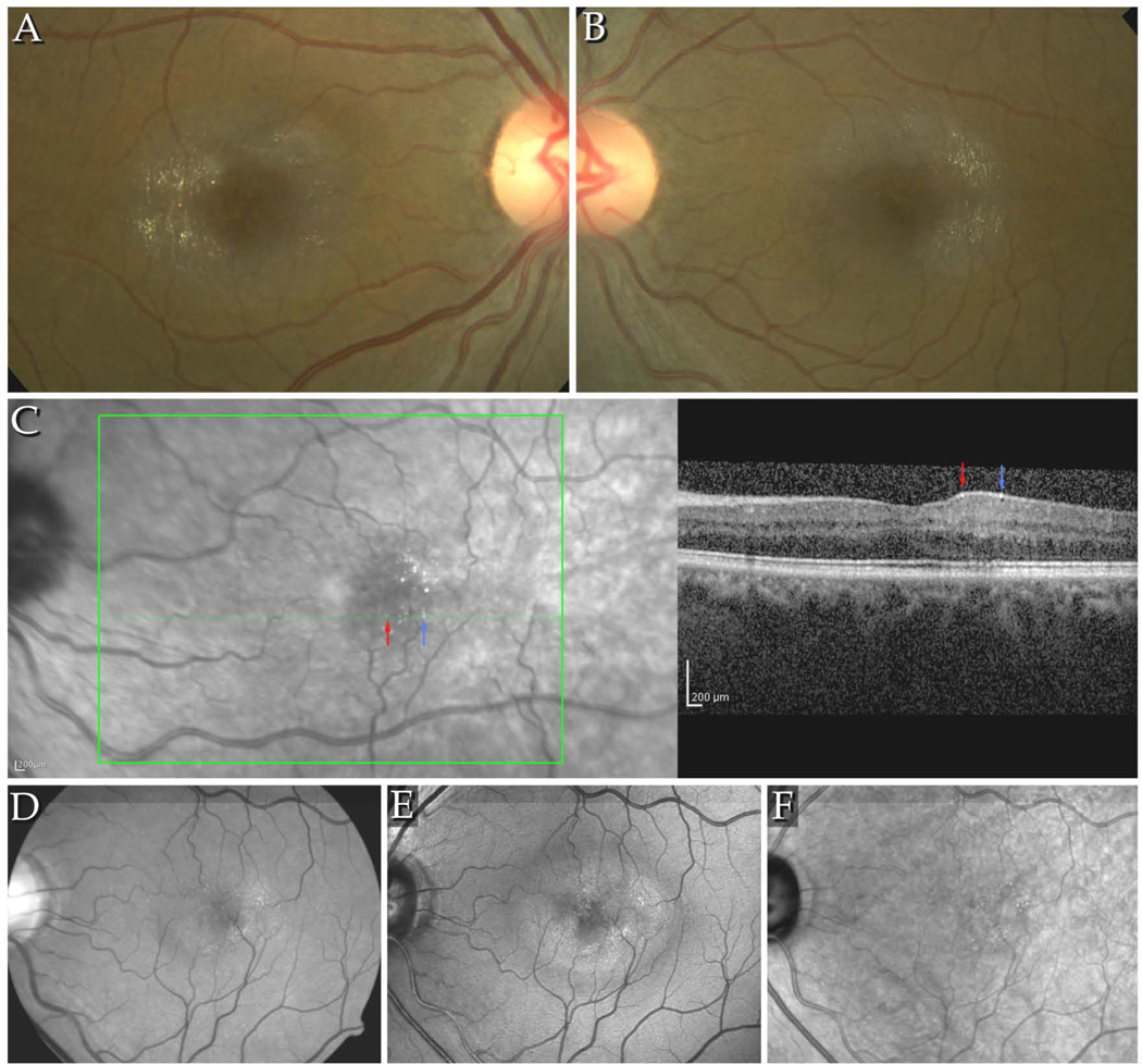
Figure 1.
A–B Colour fundus images of the right and left eyes of a patient with signs of stage 3 MacTel, including retinal crystals. The crystals are arranged in a pattern along the fibres of the NFL, within a round area with an approximate diameter of up to 4000µm, centred on the fovea. Typically, few crystals are seen at and immediately around the fovea. The area temporal of the fovea, corresponding to the raphe, contains also usually less crystals. C. IR+OCT image combination from the Heidelberg Spectralis. Red and blue arrows indicate identical retinal crystals in the IR image and the B-scan. The crystals appear to be located to the NFL. D–F. Detectability of crystals in different imaging modalities: D shows a red-free image taken with a fundus camera, E and F: confocal blue light reflectance (CBR) and infrared images (Heidelberg Spectralis HRA+OCT). Best detectability of crystals is provided by the CBR images. Crystals were located within the area of increased reflectivity in CBR.

