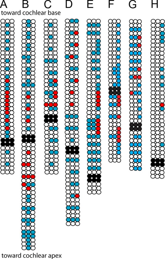
Figure 7.

Receptive field maps for eight type II afferent dendrites. Three rows of circles indicate three rows of OHCs. Black circles: OHCs removed to expose dendrites at recording site. White circles: unstimulated OHCs. Blue circles: stimulated OHCs that did not evoke postsynaptic EPSCs. Red circles: stimulated OHCs that evoked EPSCs in the postsynaptic type II afferent. A–D, 1 s duration puffs evoked multiple EPSCs from OHCs indicated by red circles. Due to tissue orientation, OHC row number could not be determined in experiment in A. E, 100 or 10 ms duration puffs of high potassium solution evoked EPSCs from some OHCs. F–H, 10 ms puffs of high potassium solution were used to evoke synaptic release from OHCs.
