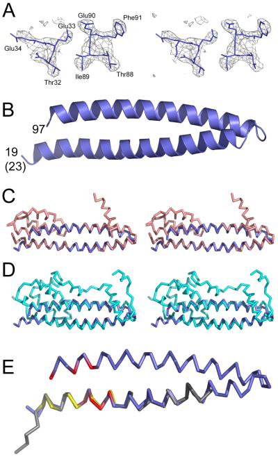
Figure 2.
Crystal structure of the helical hairpin of CHMP4B.
(A) Stereo image of the electron density map calculated based on the SAD phases without density modification.
(B) Ribbon diagram of the CHMP4B helical hairpin containing residues 23-97. Note that the crystallized construct contained four extra residues at the N-terminus, which are in a helical conformation.
(C) Stereo images of CHMP4B (blue) and CHMP3 (salmon) (Protein Data bank (PDB) ID 3FRT) based on superpositioning of the Cα atoms.
(D) Stereo images of CHMP4B (blue) and IST1 (cyan) (PDB ID 3FRR) based on superpositioning of the Cα atoms.
(E) Superpositioning of the Cα atoms of CHMP4B (blue) and Vps20 (gray) (CHMP6) (PDB ID 3FTU; the CHMP4B residues affecting CC2D1A interaction are labeled in red and the Vps20 residues involved in ESCRT-II Vps25 interaction are shown in yellow.

