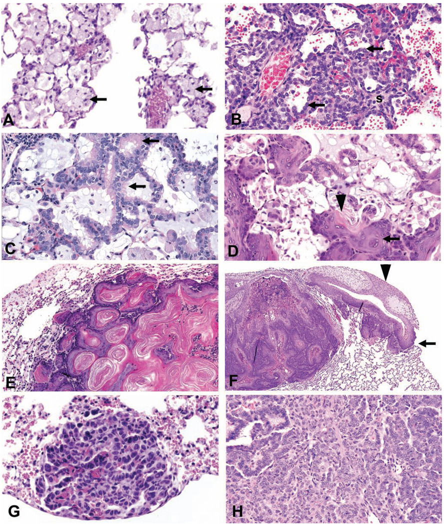
FIGURE 2.
Lung. (A) Alveolar histiocytosis in a rat characterized by the accumulation of macrophages (arrows) with abundant foamy cytoplasm in alveoli. (B) Alveolar epithelial hyperplasia in a rat characterized by an increase in cuboidal type II cells (arrows) lining interalveolar septa (s) which are thickened. (C) Bronchiolar metaplasia of alveolar epithelium in a rat where alveolar type I cells are replaced by columnar ciliated cells (arrows). The alveolar lumen contains a basophilic mucin-like material. (D) Squamous metaplasia in a rat lung characterized by the metaplasia of alveolar epithelial cells to squamous cells (arrow), distortion but not obliteration of the normal architecture, and keratin (arrowhead) formation. (E) Benign cystic keratinizing epithelioma in a rat lung characterized by a complex, thick irregular wall, expansive growth into contiguous alveoli, lack of orderly maturation, and the formation of a large keratin-filled cavity. (F) Squamous cell carcinoma in a rat lung characterized by extension through the pleura (arrow) and scirrhous reaction (arrowhead). (G) A/B adenoma in a mouse lung consisting of cuboidal to polygonal cells in a papillary pattern with a distinct border and mild compression. (H) A/B carcinoma in a mouse lung consisting of anaplastic cells with a heterogeneous growth pattern. All sections were stained with hematoxylin and eosin.

