
Figure 3.
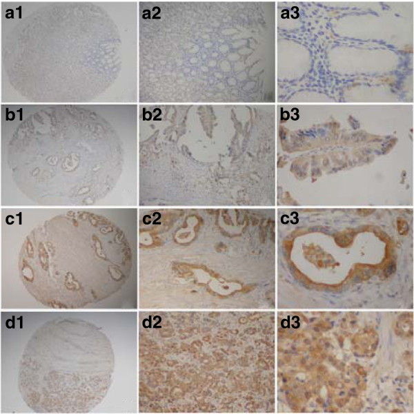
Immunohistochemical staining for S100A6 in gastric cancer lesions and noncancerous tissues.a, 1 to 3, S100A6 negative in noncancerous tissues; magnifications were × 40, ×100, and × 400, respectively. b, 1 to 3, S100A6 was highly expressed in well differentiated adenocarcinoma; magnifications were × 40, ×100 and × 400, respectively. c, 1 to 3, S100A6 was highly expressed in moderately differentiated adenocarcinoma; magnifications were × 40, ×100 and × 400, respectively. d, 1 to 3,S100A6 was highly expressed in poorly differentiated adenocarcinoma; magnifications were × 40, ×100, and × 400,respectively.