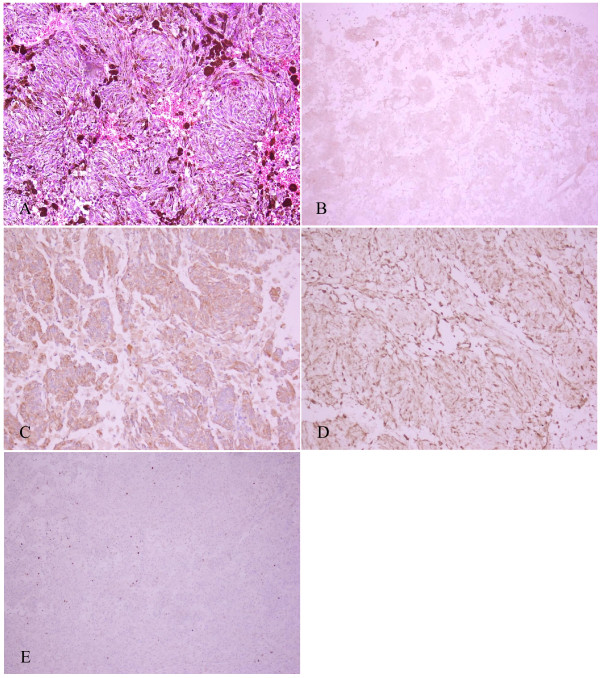
Figure 3.
Histopathological characteristics of the tumor. A: Melanin pigment is abundant and the cells are arranged in bundles with prominent nuclei. (H&E, 200×). B: HMB-45 (+) staining of tumor cells. C: Vimentin (+) staining of tumor cells. D: S-100 (+) staining of tumor cells. E: Ki-67(+), less than 1%.

