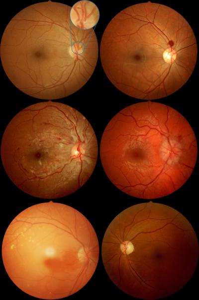
Figure.
Examples of non-mydriatic fundus photographs obtained by nurse practitioners during the FOTO-ED study. Top left: Normal posterior pole, showing the normal field of view of non-mydriatic fundus photography, which includes the optic nerve, macula, and major retinal vessels. The enlarged inset compares the single field of view of the most commonly used conventional direct ophthalmoscope which only shows part of the optic nerve head, and requires active exploration of the fundus by the examiner. Top right: Intraocular hemorrhage. Middle left: Grade IV hypertensive retinopathy with optic nerve edema, arterial attenuation, and retinal hemorrhages. Middle right: Optic nerve edema from intracranial hypertension. Bottom left: Acute retinal ischemia from central retinal artery occlusion. Bottom right: Optic nerve pallor. The black backgrounds of the original images were cropped and the brightness/contrast was adjusted.

